A clinical examination was performed to evaluate the pigmentation of the maxillary gingiva.
Assessment Included:
Clinical intraoral examination
Photographic documentation
Intraoral Images: — (Before-treatment images)
Consultation & Advanced Diagnostics
The patient’s gingival condition was reviewed, and the findings were documented to finalize the treatment approach.
Diagnostics Used:
Clinical intraoral examination
Photographic analysis
Additional Records:
Intraoral photographs
Extraoral photographs
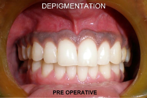
Clinical Findings:
Melanin pigmentation was seen in the maxillary gingiva. The gingival tissues appeared healthy.
Intraoral Images: — (Before-treatment images)
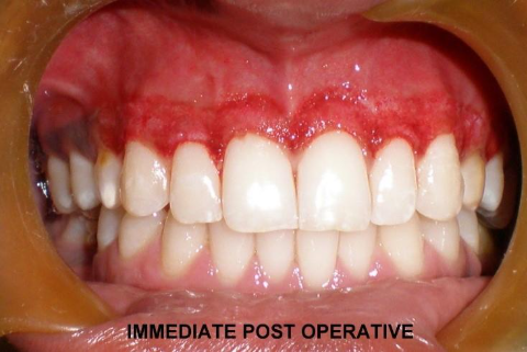
Post-Treatment Care
Medications: A short course of analgesics was prescribed for recovery
Oral Hygiene: Instructions were provided to maintain cleanliness of the treated area
Post-Treatment Instructions
Avoid hot food and drinks for 1–2 days
Do not brush or floss the treated area for a few days
Visit the clinic after one week for review
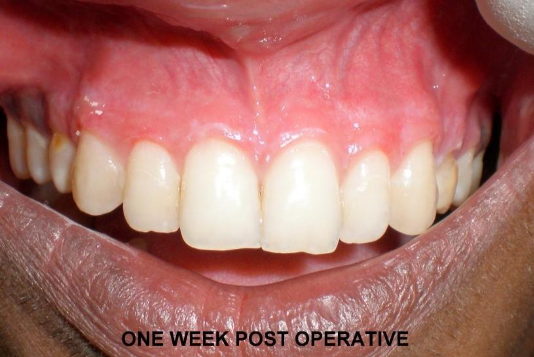
After Treatment (The Result)
At the one-week follow-up, the gums were even in color and pink in appearance.
After-treatment images
Patient Feedback
“I was worried about the progressive darkening of my gums. After the gum depigmentation treatment with Dr. Sajith Abdul Lathif, my gums are now even in color and pink, which makes my smile look much better. I am thankful to Dr. Sajith Abdul Lathif and his team for their care.”