Skip to content Skip to footer

Root Canal Treatment in a Lower Molar with Radix Entomolaris

Patient Clinical Profile

  • Gender: — Male
  • Age: — 28 Years

Issues Faced by the Patient

The patient consulted at Al Salam Health with the following concerns:

• Tooth Pain:

Severe pain in the right lower back tooth for the past three days, occurring after taking hot food and drinks and continuing for about 2–3 hours.

Before Treatment (Initial Assessment)

A clinical examination assessed the condition of the affected tooth.

Assessment Included:

  • Clinical dental examination
  • Radiographic evaluation (periapical radiograph – IOPA)

Radiographic Assessment

A diagnostic radiograph evaluated the internal structure and surrounding tissues.

Radiographic Findings:

A diagnostic radiograph showed secondary distal caries extending close to the pulp (inner nerve area), along with periapical changes and widening of the lamina dura (increased space around the root).

Radiographic Image: — (Pre-treatment radiograph)

Treatment Procedure

Access Opening & Canal Identification
  • Access cavity preparation was performed under local anesthesia
  • The distal canal was initially located towards the buccal side, indicating the presence of an additional canal on the lingual side (radix entomolaris)
  • The access cavity was modified from a triangular to a trapezoidal shape to facilitate location of the fourth canal
Canal Identification & Working Length Determination
  • Canal orifices were located using a DG-16 endodontic explorer
  • Canal patency was established using a #15 K-file (Mani, Japan)
  • Working length was determined using an apex locator (Root ZX, J. Morita) and reconfirmed radiographically

The treatment was provided under the care of Dr. Sumeera K Hussain, G.P Dentist, with 10+ years of clinical experience.

Treatment Image: — Working length determination radiograph

Cleaning and Shaping

  • Biomechanical preparation was completed using rotary endodontic files (EdgeEndo system)
  • Irrigation was carried out using 2.5% sodium hypochlorite during instrumentation
  • Final irrigation was performed using 17% EDTA

Obturation & Restoration

  • Obturation was performed using gutta-percha points with the lateral condensation technique
  • The access cavity was restored using composite resin (Tetric N-Ceram, Ivoclar Vivadent)
  • A post-obturation radiograph was taken

Treatment Images: — Master cone radiograph / Post-obturation radiograph

After Treatment (Follow-up Review)

At the 6-month follow-up, the patient was asymptomatic (no pain, discomfort, or swelling). Radiographic evaluation showed no signs of periapical pathology.

Post-operative Image: — Follow-up radiograph (6 months)

Clinical Insight

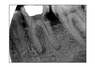
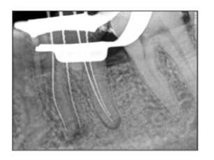
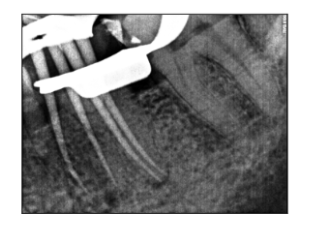
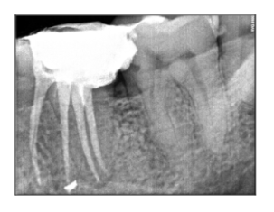
The complexity of the root canal system often poses a challenge to the clinician. Failure to identify the RE can affect the prognosis of endodontic treatment. Thus, accurate diagnosis and a thorough understanding of variations in root canal morphology, prevalence, and canal configuration of radix entomolaris are prerequisites for endodontic success.

Patient Feedback

“I had severe pain and was worried about the treatment. Dr. Sumeera K Hussain clearly explained the condition and the procedure. The treatment was comfortable, and I didn’t experience any major discomfort. I’m thankful to Dr. Sumeera and the team at Al Salam Health for their care and support.”

Subscribe for the updates!

[mc4wp_form id="461" element_id="style-11"]