Skip to content Skip to footer

Open Apex Treatment in a Traumatized Front Tooth Using MTA Apical Plug

Patient Clinical Profile

  • Gender: — Male
  • Age: — 21 Years

Issues Faced by the Patient

The patient consulted at Al Salam Health with the following concerns:

• Tooth Damage & Discolorationn:

Broken upper front tooth with visible discoloration

• History of Trauma:

Dental trauma reported approximately 9 years ago

Attending Doctor: Dr. Sumeera K Hussain

Before Treatment (Radiographic Assessment)

An initial radiographic evaluation was carried out to assess the condition of the affected tooth.

Assessment Included:

  • Periapical radiographic examination

Radiographic Findings:

  • Incomplete root formation (open apex) in tooth #9

  • Presence of a periapical lesion at the root apex

Radiographic Image: — (Pre-treatment radiograph)

Consultation & Advanced Diagnostics

During the consultation, the patient’s dental condition was evaluated and appropriate treatment was discussed.

Diagnostics Used:

  • Percussion test (horizontal & vertical)
  • Pulp vitality test using Endo Frost

Clinical Findings:

The examination showed the following:

  • Pulpal exposure present
  • No tenderness on percussion (both horizontal and vertical)
  • No history of swelling, fever, night pain, or draining sinus
  • No abnormal mobility detected
  • Negative response to cold vitality test (Endo Frost), suggestive of pulp necrosis

Diagnosis:

  • Non-vital tooth with open apex and associated periapical pathology

Treatment Decision:

  • Considering the wide open apex, MTA apical plug was decided as the treatment plan.

  • The treatment plan was explained to the patient, and informed consent was obtained.

Treatment Procedure

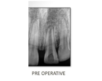
Phase 1: Access Opening and Canal Shaping
  • Coronal access cavity was prepared using a round bur
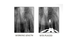
  • Working length was determined using a #80 K-file with radiographic confirmation
  • Cleaning and shaping was done using crown-down instrumentation
  • Irrigation was carried out with 2.5% sodium hypochlorite (NaOCl) with controlled aspiration
Phase 2: Intracanal Medication
  • The canal was dried using sterile paper points
  • Calcium hydroxide (Ca(OH)₂) mixed with saline was placed as an intracanal medicament
  • The tooth was temporarily restored

The patient was recalled after one week

Phase 3: MTA Apical Plug Placement
  • The intracanal medicament was removed
  • The canal was irrigated with NaOCl and dried using paper points
  • Mineral Trioxide Aggregate (MTA – Angelus) was mixed according to manufacturer instructions
  • MTA was placed into the canal using an MTA gun
  • A 4 mm apical plug was formed and gently condensed using hand pluggers at the apical foramen
  • Placement was verified with radiographic confirmation
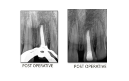
Phase 4: Canal Obturation & Restoration
  • A moistened cotton pellet was placed at the canal orifice, and the patient was recalled the next day
  • After confirming the MTA had set, the remaining canal was sealed using warm vertical compaction

The patient was referred for prosthetic crown restoration

The treatment was provided under the care of Dr. Farijan Abdul Latheef, G.P Dentist, with 10+ years of clinical experience.

Treatment Progress Images

Final Evaluation

The results were reviewed using clinical and radiographic assessment.

Evaluation Included:

  • Healing of the periapical
  • Progress in apical closure
  • No post-operative symptoms

Post-Treatment Result

At 6-month review, the patient showed:

  • No post-operative pain or discomfort
  • Healing at the apex
  • Apical closure achieved
  • Tooth remained firm and comfortable

After-treatment images

Patient Feedback

“I had a broken front tooth for years after an old injury and didn’t know it could be treated this well. The procedure was very comfortable, and I’m relieved there’s no pain now. Thank you to the doctor, Dr. Sumeera K Hussain, and the team for the care.”

Subscribe for the updates!

[mc4wp_form id="461" element_id="style-11"]